Facilities
The neuroimaging resources at Wayne State University are world class with several state-of-the-art MRI and NMR scanners for human and animal studies. Listed below are the primary, research dedicated systems used by members of the BRAIN division.
Magnetic Resonance Research Facility
 The MR Research Facility (MRRF) is committed to the development of MR methods and their application in preclinical and clinical populations to better understand the nature of brain disorders and diseases. The MRRF promotes the use of MR-based methods to the WSU scientific community and supports the implementation of MR methods through education, assistance in experimental design, and data collection and analysis.
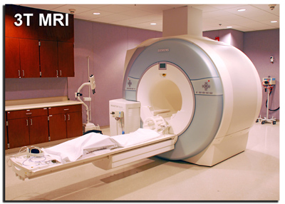
The MRRF center is equipped with a 100% research dedicated, high-field 3 Tesla Siemens Verio whole-body human MRI scanner capable of collecting high spacial resolution anatomical MRI imagages, functional MRI or fMRI data, diffusion tensor imaging or DTI data and multi-nuclear spectroscopy or MRS data (1H and 31P) as well as echo-planar spectroscopic imaging (EPSI) data. This system also has an industry leading 32-channel head coil for superior image quality.
The MRRF center is located in the Harper University Hospital, department of Radiology and is directed by E. Mark Haacke.
Pediatric Imaging Center
Two MRI systems dedicated to scanning children are available for research at the Children's Hospital of Michigan, Department of Pediatric Imaging. This includes a high-field 3 Tesla short-bore MRI system as well as a 1.5 Tesla system capable of conducting anatomical MRI, fMRI, DTI and 1H MRS experiments. Both instruments have an 8-channel head coil for parallel imaging.
Small Animal MRI Facility
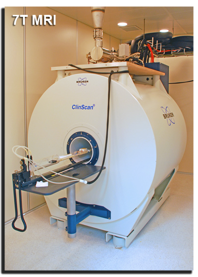 The Small Animal MRI Facility is equipped with a 7 Tesla ClinScan animal MRI scanner capable of measuring tissue structure and function at the level of microns. This system is designed to further facilitate translational research from 'mice to human' in the field of preclinical and molecular imaging by using a Siemens' interface that allows a direct and fast transfer of preclinical studies on animal models to clinical studies on humans.
The 7 Tesla high-field MRI system is located in the Elliman Clinical Research Building and is directed by Bruce Berkowitz.
Molecular Neuroimaging Laboratory
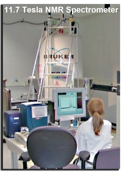 The Molecular Neuroimaging Laboratory, directed by Dr. Galloway, is located at Gordon Scott Hall and is equipped with a high-resolution, 11.7 Tesla vertical bore Bruker microimaging system. The system is capable of collecting not only multi-nuclear NMR spectra at an incredible spectral resolution (i.e., 1H, 2H, 7Li, 13C, 17O, 19F and/or 31P NMR) but also MRI images of small animals or extracted tissue. High resolution magic angle spinning or HRMAS is another technique that has proven to be a highly effective in profiling the chemical composition of brain tissue and gaining a greater understanding of drug or environmental effects on the brain biochemistry.